Thursday, June 28, 2012

Inarticulate Brachiopod Larvae

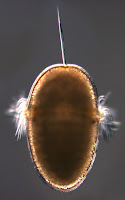
This June I participated in an oceanographic research cruise to Barbados. One of the goals of this expedition was to collect planktonic larvae of benthic marine invertebrates using a MOCNESS device. Among other things, we found larvae of inarticulate brachiopods (phylum Brachiopoda; class Inarticulata) pictured here. Brachiopods resemble bivalve molluscs (e.g. clams and mussels), but have dorsal and ventral valves rather than left and right. The valves are held together with muscles in inarticulate brachiopods, while those of articulate brachiopods are hinged. Brachiopods, as a phylum, are also characterized by the lophophore (a crown of tentacles surrounding the mouth). We collected two different kinds of inarticulate brachiopod larvae.

Larvae of the first kind were collected from relatively shallow depths (900-0 m). In this image, one can see the large circular larval shell valves, as well as the embryonic shell (the half circle at the posterior portion of the larval shell, tinged blue). The lophophoral tentacles are retracted in the first image, but extended in the second.

These tentacles form progressively during the larval life from short tentacle buds on either side of a longer unpaired anterior median tentacle (at about 12 o’clock on the second image). Consequently, the number of tentacles reflects larval age. This larva has 10 pairs of tentacles. In some species, a lophophore with this many tentacles indicates larval competence for settlement (Pennington and Stricker, 2002). One can also see that the tentacles of the lophophore are ciliated. Cilia are used for larval feeding and swimming (Rudwick 1970). This particular type of larva is produced by members of the superfamily Lingulacea which contains only two extant genera: Lingula and Glottidia (Pennington and Stricker 2002).

Larvae of another kind were collected from deeper waters (1600-900 m). Characteristically, they lack an embryonic shell, and have a pair of larval chaetae. Presence of chaetae in this larva suggests that it belongs to the inarticulate superfamily Discinacea (Pennington and Stricker 2002). Chaetae are usually thought of as a feature unique to annelid worms, but some brachiopod larvae and adults also have chaetae, which are similar structurally and developmentally to those of annelids. 

Pennington, J T and S A Stricker. 2002. Phylum Brachiopoda. In: Atlas of Marine Invertebrate Larvae. Edited by Craig M Young. Academic Press. 

Rudwick, M J S. 1970. Living and Fossil Brachiopods. Hutchinson & Co, London.

Monday, June 4, 2012

Fan worm larva

On May 10th, 2012 I waded through the calm waters of Middle Cove at Cape Arago State Park, trailing a 153μm plankton net behind me.  While sorting through the plankton I found a polychaete larva from the family Serpulidae, subfamily Spirorbinae.  Serpulids belong to an order of annelids, Sabellida, commonly known as fan worms.  Adult Spirorbins live in small calcareous tubes approximately 2-5 mm in diameter typically attached to undersides of intertidal rocks. Adults are hermaphroditic and brood embryos inside their tube, then release lecithotrophic larvae that are only briefly planktonic (Strathmann 1987).

This metatrochophore larva has a prominent ciliated band anterior to the mouth, called the prototroch, which is used for swimming. At the anterior (up) is the apical tuft, and at the posterior end there is another cilary band, called telotroch. What helped us to identify this larva is the prominent collar located posterior to the prototroch (Kupriyanova et al. 2001). The collar is the widest portion of the larval body. This larva had  5-6 segments posterior to the collar, and three pairs of ocelli. 

To my surprise, when I went to photograph the larva a few days later, I could not find it at first.  No longer content with the planktonic life, it had settled on the bottom of the culture dish and built a calcareous tube (left). The tube is smooth, non transparent, and coils to the right (dextral). The characteristics of the tube helped us to further classify this specimen as likely belonging to the genus Circeis (Blake and Ruff 2007). 

You can see on the subsequent photograph that it got quite a bit longer in 4 days. The juvenile worm is caught peaking out of it’s tube in the first picture. One can discern a radiolar crown of tentacles used for feeding and respiration (Blake and Ruff 2007), red eyespots, and an operculum.  When disturbed the animal quickly retreats back into its tube, and shuts the operculum (below).

Blake JA, and Ruff RE.  2007. Polychaeta.  In: The Light and Smith Manual:  Intertidal Invertebrates from Central California to Oregon 4th Edition.  Edited by James Carlton. University of California Press, Berkeley.  

Kupriyanova, E.K., E. Nishi, H.A. ten Hove, & A.V. Rzhavsky 2001. A review of life history in serpulimorph polychaetes: ecological and evolutionary perspectives. Oceanography and Marine Biology: an Annual Review 39:1-101.

Strathmann, Megumi F. 1987.  Phylum Annelida, class Polychaeta. In: Reproduction and Development of Marine Invertebrates of the Northern Pacific Coast.  United States: University of Washington Press. 

Sunday, June 3, 2012

Development of the sea cucumber Cucumaria miniata

This series of images illustrates the development of a sea cucumber Cucumaria miniata. I collected adults of C. miniata from Lighthouse beach in Charleston, Oregon on April 24 2012 hoping to obtain gametes from natural spawning because this species is known to reproduce from mid-March to late April. I kept the adults in a sea table with flowing sea water at ambient temperature and crossed my fingers. On April 28 2012, two males and three females spawned. The adults were separated before spawning, so I collected the gametes and fertilized the eggs. Cucumaria miniata is considered a direct-developer. Even though it has a swimming larva, called doliolaria, this larva does not feed in the plankton. In contrast, some other sea cucumbers have a feeding auricularia larva, which eventually metamorphoses into a doliolaria stage. As is typical for direct developing sea cucumbers, C. miniata has large yolky eggs (500 µm in diameter and bright green in this species). A few hours after fertilization I observed early cleavage – a four cell stage is pictured here.

The second picture shows a 60-hour old early doliolaria larva of C. miniata – the preoral lobe (right) is more opaque compared to the posterior end of the larva (left). Although these do not feed they do have a vestige of a gut. Early on the larva is uniformly ciliated, but the advanced doliolaria larva of this species swims using three transvers ciliary bands (not shown).

By day seven of development one can see there are five-primary tentacles protruding ventrally toward the anterior end of the larva (about six o'clock on the image on the left). The five primary tentacles surround the mouth. At this stage the individual is referred to as a pentactula. The pentactula of this species also has two primary podia (one clearly visible on the image to the left) emerging from little pores (called podial pits) at the posterior end.  

The bottom image shows several of the many calcareous spicules in the pentactula. After two weeks of planktonic life the larvae (as pentactulae) settle on the undersides of rocks near conspecific adults.

Sewell, M. and McEuen, F. Phylum Echinodermata: Holothuroidea. In: Atlas of Marine Invertebrate Larvae. Edited by Craig M Young. Academic Press.

McEuen, F. Phylum Echinodermata, Class Holothuroidea. In: Reproduction and Development of Marine Invertebrates of the Northern Pacific Coast. Edited by Megumi F Strathmann. University of Washington Press.

Genital plate formation in echinopluteus larva

Indirect development often involves a major change in body plan. The larva of the purple sea urchin, Strongylocentrotus purpuratus, has bilateral symmetry, and the adult has 5-fold radial symmetry. As the echinopluteus larva approaches metamorphosis clues to the body-axis shift start to appear. The first image shows a lateral view of the larva. The two vertical white lines are calcareous rods that support the arms of the larva.  They appear to glow because I used polarized light microscopy. Forming around each rod is a honeycomb-like structure which will become one of the genital plates in the adult. After metamorphosis, these plates are located on the aboral side of the adult (the side opposite the mouth) and surround the mouth. They are called genital plates because the gonads (testes or ovaries) open to the outside via little holes (called gonopores) in these plates. The endoskeleton of adult urchin, called the test, is made up of many such closely fitted calcareous plates.

The second picture is a close-up aboral view of the test of an adult urchin. Surrounding the large hole in the center (the anus) are the apical plates. You can also see five smaller openings, the gonopores, in the genital plates. Starting at 12 o’clock is the genital plate 2 or G2. This much larger plate perforated by a large number of small pores is called the madreporite; it connects the water-vascular system of the urchin to the outside. Clockwise from G2 are the other four genital plates in the following order: G3, G4, G5, and finally G1. I think the left and right genital plates on the larval image above correspond to G5 and G3 respectively (Emlet, 1985).

Emlet, R. B. 1985. Crystal Axes in Recent and Fossil Adult Echinoids Indicate Trophic Mode in Larval Development. Science 230: 937-940.

Saturday, June 2, 2012

Müller’s larva

On May 3rd and 4th 2012, I performed plankton tows at the South Cove of Cape Arago, near Charleston, OR as the tide was coming in.  I placed a 153μm plankton net into a channel of water flowing between smooth rocks. In this manner I was able to obtain interesting samples, which included, among other things, many specimens of Müller’s larva pictured here. 

Johannes Müller (1801-1858), a German physiologist and the inventor of the plankton net, first described larval forms of many phyla including Platyhelminthes, or flatworms, whose planktonic larvae retain his name (Smith et al. 2002). The term “Müller’s larva” is used to describe planktonic larvae in the Polycladida order of the class Turbellaria. Müller’s larva is ciliated and characterized by several paired and unpaired lobes (left). The lobes bear cilia that are longer than cilia on the rest of the body. At the anterior (up) and posterior (down) end of the larva there are tufts of longer cilia (apical and caudal, respectively). The apical tuft originates from the apical organ, a sensory structure that is associated with the central nervous system (Rawlinson 2010).

When I collected these larvae they were fairly uniform in size approximately 180 μm long by 80 μm wide. Their shape was simliar, with lobes present in all of the specimens.  When I observed them two weeks later some had lost their characteristic lobes. The second photo shows a larva where the lobes are present, but do not protrude much.  Müller’s larvae may also have several pairs of eyes.  The two areas of black pigmentation at the anterior portion are a cerebral eye and an epidermal eye, which is closer to the apical tuft. 

The last photo (left) shows a, presumably, more advanced larva that has completely resorbed its lobes and has begun to take the characteristic shape of an adult flatworm. In this larva you can clearly see the two cerebral eyes, with one of the epidermal eyes lower and to the right. When observed two weeks later this larva had not noticeably changed in shape or form. Little is known about the development of Müller’s larva, and it would be interesting to investigate the mechanisms driving these morphological changes.

Smith NF, Johnson KB and Young CY. 2002. Platyhelminthes. In: Atlas of Marine Invertebrate Larvae. Edited by C. M. Young. Academic Press. New York.

Rawlinson KA. 2010. Embryonic and post-embryonic development of the polyclad flatworm Maritigrella crozieri; implications for the evolution of spiralian history traits. Frontiers in Zoology. 7:12

Friday, June 1, 2012

Chiton development


This picture shows a chiton egg which I collected from the plankton on February 8th, 2012, off of a dock in Charleston, OR. Chitons are marine mollusks characterized by a shell made up of eight separate plates. Chiton shell plates washed up on the beach are often referred to as “butterfly shells.” Chiton eggs are very distinctive because they are surrounded by a thick and often ornate hull (as seen in this picture). The hull has been found to reduce the sinking rate of the egg as well as to focus sperm to specific regions on the egg surface (Buckland-Nicks, 1993) . 

This is a trochophore larva that hatched out of a chiton egg. Chiton trochophores have a long apical tuft and an equatorial ciliary band called a prototroch, which you can see on this picture. Chiton trochophores are different from any other kind of trochophore because their two eyes are located posterior to the prototroch (instead of anterior). After a few days of development these larvae begin to form shell plates. 

This picture shows seven transverse bands on the dorsal side of the larva posterior to the prototroch. These bands delineate the boundaries of the future shell plates; the eighth plate appears later in development (anterior to the prototroch). The dark band is part of the mineral skirt, which can be seen more clearly in the photo below. The foot and mouth of the chiton is developing on the ventral surface. 

I took this picture using polarized light to show the mineral spicules in the epidermis surrounding the shell (this area is called the girdle) and the seven initial shell plates which are beginning to form.   

Buckland-Nicks, J. 1993. Hull capsules of chiton eggs: parachute structures and sperm-focusing devices? Biological Bulletin. 184: 269-276.