Saturday, April 28, 2012

Viviparous brittle-star Amphipholis squamata

Amphipholis squamata are simultaneous hermaphrodites, which means they produce both eggs and sperm at the same time, and they self-fertilize. The exciting part is that this tiny brittle star is also viviparous. We dissected adults in class to find brooded embryos.

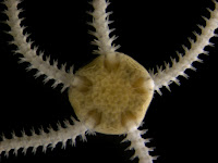
Amphipholis squamata oral wholeThe long slender arms are made up of a series of flexible joints and can bend and coil. This brittle star will unexpectedly fold up to conceal itself or reach out quickly to make a sneaky escape. In preparation for these photos, I  immersed the brittle star in a solution of MgCl2 to anesthetize it. It almost immediately relaxed and stayed still through the subsequent dissection.

Amphipholis squamata aboral detailA close view of the aboral side shows the tiny central disc of an adult (4 mm in diameter). The disc isn't strictly circular but pentagonal. The whole body has a five-fold symmetry so there are five (or multiples of five) of every structure, as is characteristic of the phylum Echinodermata, in general.

Amphipholis squamata oral detailA close view of the oral side shows a smaller pentagon at the center. This is made up of five triangular teeth which, when open, reveal the mouth. On either side of each arm are dark lines. These are bursal slits, entrances to ten sac-like spaces called genital bursae. 
Ovaries and testes flanking the bursae release sperm and eggs into the bursae through gonopores. Fertilization happens internally and the embryos develop and grow inside the parent's body. The juvenile emerges from the bursal slit when mature. Amphipholis squamata is fertile year-round and is able to brood multiple young at different stages of development.

Amphipholis squamata juvenile detailUsing sharp forceps, I searched for brooded embryos by pulling back the tissue between two bursal slits. After my third try I was pleased to find this tiny juvenile. See also a post by Amanda Clark which shows two dissected juveniles of A. squamata at different stages of development.

Friday, April 27, 2012

Field Trip to South Cove

The Pacific Northwest is a particularly rich location to experience the biodiversity of marine life.  Spring is when many species reproduce making it an excellent time of the year to study embryonic and larval development of marine invertebrates. A point of interest is the South Cove of the Cape Arago headland as it offers students here at the OIMB an opporunity to observe and study a diverse array of organisms.  On April 24 at 8:30 a.m., with our gear in tow, we left for the South Cove to catch the -0.3 foot low tide at 9 a.m.  By van we made our way six miles south of the OIMB campus down Cape Arago highway to look for particular species of brooding bryozoans. 

Sticking close to more experienced students and our instructor Svetlana, I saw many interesting species including this morning sunstar, Solaster dawsoni, being held up by my classmate Ashley (left). Other common species of the South Cove intertidal zone we observed include the worlds largest chiton, Cryptochiton stelleri, commonly known as the gumboot chiton, the worlds largest sea star, Pycnopodia helianthoides, commonly known as the sunflower seastar, and a model organism for developmental biologists, Strongylocentrotus purpuratus, commonly known as the purple sea urchin, to name a few. After two hours in the intertidal, we headed back to the lab to study our specimens. In addition to finding the brooding bryozoans we were looking for, we found many other interesting species that we will study in class. 

A rather noteworthy member of this menagerie pictured in the center of the glass dish is the veiled chiton, Placiphorella velata. It is a rare find because this species typically occures in the very low intertidal or subtidal. Unlike most chitons which graze on algae, this predatory chiton waits for small crustaceans and worms to wander under its veil and rapidly (less than a second) lowers the hood to trap its prey. It then swallows the smaller bits whole or uses its radula to scrape and break apart parts of larger prey.

Identifying pinnotherid crab larvae

The first picture is of a larva of a crab from the family Pinnotheridae collected from the plankton on April 19th, 2012 from the boat basin in Charleston, Oregon.  Pinnotherids, commonly known as pea crabs, are crabs that live commensally with a variety of organisms e.g. mollusks, annelids and echinoderms.  This zoea has relatively short rostral and dorsal spines in addition to two lateral spines. Studies have shown that lateral spines likely serve to reduce predation by planktivorous fishes. Additionally, one can see one of the two large compound eyes of this larva.Pea crab zoeas are some of the most ubiquitous zoea larvae encountered in plankton tows in this part of the world.  However, zoea larvae of many brachyuran crabs are very similar and identification can be tricky. The two bottom pictures highlight the differences between zoea larvae of a pea crab and a crab from the family Cancridae.

In the second picture we see a close up of the telson (terminal segment) and the two preceding abdominal segments excised from a pea crab zoea. The enlarged last abdominal segment (wider than the telson) that you can clearly see on this picture is a the identifying feature of pea crabs in the Northeast Pacific.  Furthermore, most pinnotherid zoeas have small narrow telson.

In the third picture we see the telson and last four abdominal segments of a zoea from the genus Cancer.  One can see a pair of spines on each of the abdominal segments, however none of the segments is expanded to the point of being wider than the telson.  Additionally, the telson  of a cancrid zoea is wider than that of the pinnotherid zoea and is forked.

Thursday, April 26, 2012

Coronate larva of Crisia sp.


This picture shows a fragment of a bryozoan colony Crisia sp. I collected it at South Cove just south of Charleston, OR during low tide in April 2012. The colonies of Crisia sp. look like upright branching whitish tufts about 1.5 cm tall on the underside of rocks and under rock ledges. One can see openings of many individual feeding zooids (autozooids) and one zooid specialized for reproduction (gonozooid). Gonozooid is larger than the autozooids and looks like a yellow “pouch”. Crisia is interesting because it exhibits an unusual reproductive strategy called polyembryony. A single zygote is initially deposited within the gonozooid, it receives nutrients from the mother zooid via a kind of placenta, grows, and buds off secondary embryos, which, in turn, can bud off tertiary embryos. In this way each gonozooid ends up  filled with many genetically identical small embryos - a kind of embryonic cloning.  The calcareous wall,  which protects the gonozooid (much like the other zooids in the colony), is clearly visible.

The second picture shows a gonozooid in which I cracked and removed part of the wall with a pair of sharp forceps to expose the embryos. Each of the small yellow spheres is a brooded embryo. These embryos are tightly packed inside the gonozooid and readily spill out when it is opened. These brooded embryos emerge as ciliated coronate larvae.


The bottom image is a side view of a coronate larva I removed from a gonozooid of Crisia. Most of the larval surface is covered by a ciliated epithelium called corona ciliata. Corona ciliata is used for locomotion. Coronate larvae do not have a shell (unlike the other kinds of bryozoan larvae (cyphonautes and pseudocyphonautes), or a gut (so they do not feed). They spend only a few hours in the plankton before settling. Coronate larvae of Crisia are anatomically very simple. Aside from the corona ciliata they have an internal sac (not visible here) and an aboral non-ciliated region clearly visible here (top). A portion of this non-ciliated region is invaginated in a circular groove, which corresponds to pallial epithelium.   The coronate larva of Crisia sp. lacks the pyriform organ and vibratile plume found in coronate larvae of some other bryozoan species (e.g. Bugula and Schizoporella).

Nectochaete larva of Harmothoe sp.

This polycheate nectochaete larva was found in a plankton tow collected in the Charleston, OR boat basin. Using a plankton identification guide (Crumrine 2001) with a dichotomous key I identified this larva as belonging to the genus Harmothoe (Family Polynoidae, commonly known as scale worms). This larva has nine setigers (segments bearing setae) and five pairs of elytra (scales that characterize the worms of this family).

The first picture was taken on the day when the larva was collected – April 12, 2012. It is a dorsal view, so one can clearly see the five pairs of large scales (elytra) running along the anterior-posterior axis. The third pair of elytrae is unpigmented, whereas the rest of them have a pigmented margin (appears golden in dark field). The larva has three pairs of large ocelli (two of which are in focus). There are also three pointed antennae on the anterior-most segment (called prostomium), all tinted with the same golden pigment as the elytrae.

The second picture was taken six days later. This is a ventral view, so one can clearly see the nine pairs of parapodia (appendages) with chaete (setae), the two ventral palps with bulbous base – one on each side of the mouth, and the two anal cirri (appendages on the posterior-most segment, called the pygidium). Worms in the genus Harmothoe either brood early embryos or spawn (release eggs and sperm) directly into the plankton. I hope to continue to follow this larva through its development during the course and try to determine which species it belongs to.

Crumrine, L. 2001. Polychaeta. In: An Identification Guide to the Larval Marine Invertebrates of the Pacific Northwest. Edited by Alan Shanks. OSU Press, Corvallis.

Unidentified actinotroch larva

Our class collected plankton on April 12, 2012 using a 153-μm mesh size net towed behind a boat two miles outside Coos Bay, OR. The two pictures show the 0.7 mm long unidentified actinotroch larva of a phoronid worm that I found in this sample.

The top image is focused on the tentacles, each with a single black pigment spot. This larva had 14 tentacles total. At the anterior end (up) one can see the pre-oral hood, and at the posterior - the telotroch, a prominent ciliary band that surrounds the anus and propels the larva. The bottom picture shows the same individual, but focusing deeper in to show the internal structures. At the apex of the pre-oral hood, one can see a thickened region of epithelium - this is the apical sense organ (the larval brain). It may play a role in substrate selection during metamorphosis (Johnson and Zimmer, 2002).

On this picture one see almost the entire digestive tract. The oral hood encloses a funnel-shaped vestibule, at the tip of which is the mouth. The mouth leads into the stomach (a large oval shape that occupies the majority of space in the larval trunk). The stomach connects to the hindgut - a short straight tube which opens at the posterior end via an anus.One can also see a small but distinct mid-ventral (to the right on the picture) invagination just posterior to the tentacles (and next to a pigment granule). It is called the metasomal sac. This sac will grow throughout larval development and eventually wrap around the larval gut. At the onset of metamorphosis the metasomal sac is everted to form the trunk of the adult worm. The entire larval gut is pulled into the everted sac, thus shortening the larval axis, and bringing the mouth and anus closer together to form the U-shaped adult gut.

Johnson, KB and Zimmer, RL. 2002. Phoronida p.430. In: Atlas of Marine Invertebrate Larvae. Edited by C. M. Young. Academic Press. New York.

Phyllodocid metatrochophore

Polychaete metatrochophore larvae like this one were collected in plankton tows off the docks in Charleston, OR in April.  I identified them as members of the family Phyllodocidae, largely due to the presence of their leaf-like dorsal cirri.  Cirri are fleshy projections found on polychaete segments (e.g. prostomium, peristomium or pygidium) or their parapodia (appendages bearing chaetae).  Phyllodocid polychaetes have rounded dorsal (and sometimes ventral) cirri on each parapodium.  The cirri of this metatrochophore were particularly leaf-like in their shape and texture, which is a characteristic of the genus Phyllodoce (Crumrine 2001).  This image is a lateral view (anterior up) showing the distinct dorsal cirri, one of the two red eye spots, and the prototroch (a transverse ciliated band encircling the larva at its widest point).  One can also see the four pairs of long finger-like tentacular cirri just posterior to the prototroch.  The second image was taken at a higher magnification to show the leaf-like characteristics of the dorsal cirri.

I kept one of these metatrochophores in a sea table in our lab. When I checked in on it several days after collection, I discovered that it had grown into a juvenile worm and was crawling around on the bottom of the bowl.  At first glance this juvenile looks rather different from the metatrochophore in the above photographs.  But with closer examination, one can see the foliaceous dorsal cirri and the four pairs of long tentacular cirri just posterior to the oval prostomium (anterior-most segment bearing eyes).  The characteristics of both the metatrochophore and the juvenile suggest that this specimen belongs to the genus Phyllodoce.  Identification to species requires examination of the polychaete’s proboscis (the large semi-transparent shape that can be seen through the body wall in this picture).  The arrangement of papillae on the surface of the everted proboscis is a species-specific characteristic within this genus (Carlton 2007). But unfortunately this worm did not evert its proboscis while I was watching, and so the species remains undetermined.   

Crumrine, L.  2001. Polychaeta. In: An Identification Guide to the Larval Marine Invertebrates of the Pacific Northwest. Edited by Alan Shanks.  OSU Press, Corvallis.

Carlton, J T.  2007.  The Light and Smith Manual:  Intertidal Invertebrates from Central California to Oregon 4th Edition.  University of California Press, Berkeley. 

Brooded embryos of bryozoan Dendrobaenia lichenoides

The first image depicts a fragment of a colony of the bryozoan Dendrobaenia lichenoides with brooded embryos (pink).  The D. lichenoides specimens were gathered by hand in South Cove at Cape Arago State Park south of Charleston, Oregon on April 24, 2012.  Each globular ovicell (a calcified brood chamber attached to the maternal zooid) houses a single large embryo.  Ovicells tend to be in the center of the colony where the older zooids are located.  The embryos undergo cleavage and gastrulation within the ovicell, developing into a lecithotrophic coronate larva that is eventually released into plankton and swims briefly before settling and undergoing metamorphosis.  The seasonal timing of larval release is largely dependent upon whether the colony is over-wintering or nascent (Strathmann 1987).  

The second picture shows three ovicells with embryos that I dissected from the colony using a pair of sharp forceps.  With care and practice one can gently crack the ovicell and extract the embryo. 

The bottom picture shows an embryo I was able to dissect out of the ovicell intact.  The embryo is enclosed by a transparent egg envelope, or chorion.  This particular embryo is at a relatively early stage of development, and one can clearly see the outlines of individual cells (blastomeres).  This picture illustrates the biradial cleavage pattern characteristic of bryozoans in general.

Strathmann, Megumi F.  Reproduction and Development of Marine Invertebrates of the Northern Pacific Coast.  United States: University of Washington Press (1987): pp. 505.  Print.

Saturday, April 21, 2012

Echinopluteus

These pictures show an echinopluteus larva of the common Pacific coast species - the sea urchin Strongylocentrotus purpuratus (the purple sea urchin). I found this larva in a plankton sample collected off the docks in Charleston, OR on February 8th, 2012. All three pictures show the same specimen at the same developmental stage, but different focal planes, to emphasize different structures. The calcareous spicules making up the larval skeleton are clearly visible in the top photograph and appear rainbow-colored due to the use of polarized light. These spicules characterize the pluteus larva (found in echinoids and ophiuroids, a.k.a. sea urchins and brittle stars). The morphology of the larval skeleton is used to identify larval echinoids (sea urchins and sand dollars). The spicules support the larval arms (4 in this larva at this stage), which in turn support the ciliated band, used for feeding and locomotion. The longer the arms, the longer the ciliated band, the more efficient the larva can feed.

The middle photograph shows the mouth - the large oval shape at the anterior end of the larva (up) and a portion of the tri-partite gut, which characterizes all feeding echinoderm larvae. One can see the round stomach (in the center of the larva) and the esophagus (a short muscular tube that connects the mouth to the stomach). One can also see the two coelomic sacks (one on either side of the esophagus), which will form the body cavity and the water-vascular system of the adult urchin. The bottom photograph shows red pigment cells in the epidermis of the larva. These have been observed to function in wound healing in echinoid larvae: pigment cells in vicinity of the wound migrate to the damaged area and engulf cellular debris (George von Dassow, personal communication).

Thursday, April 19, 2012

Encapsulated Mollusk Trochophore

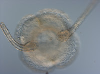
This is a trochophore larva of an unidentified gastropod mollusk collected from the plankton on February 1st 2012 off a dock in Charleston. It swims about within its remarkable bilayered egg capsule. What appears as an opening at about one o’clock is most likely the micropyle (a remnant of the oocyte’s attachment to the ovary, which is also likely the site of sperm entry). The trochophore is characterized by the presence of the prototroch - a pre-oral ciliated band, which you can clearly see in this picture. Some mollusks with intracapsular development (as opposed to pelagic development) may have a reduced trochophore stage or no trochophore stage at all. Also, the stage at which the larva hatches out of its capsule can vary from species to species: some may hatch out as trochophores, while others hatch out as veligers, as this one did.

  Molluscan veliger larvae are characterized by the presence of a shell and a velum whose ciliated lobes are used for locomotion and food capture. The velum is derived from the prototroch. The picture on the left shows a large bilobed velum of a different gastropod veliger.

Unidentified polychaete metatrochophore

This is a metatrochophore larva of an unidentified polychaete annelid that I collected from a January 2012 plankton tow off the docks in Charleston. Its gut contains a large number of yolk droplets (of various color and size). Yolk droplets of this kind are often found in eggs of annelids with lecithotrophic larvae (meaning they do not feed and derive nutrition from maternal supplies), e.g. in larvae from the family Nereidae.  This suggests that this is likely a lecithotrophic larva.  One can also see two ciliary bands. The broad ciliary band just posterior to the ocelli (eyes) is called the prototroch. The narrow band near the posterior end is called the telotroch. These are used for locomotion. In planktotrophic (feeding) larvae the prototroch may also be used for collecting food particles. This larva has three setigers (segments that bear setae), which is how we know it is an annelid.

Thursday, April 12, 2012

Micronereis nechtochaete larva


This polychaete nechtochaete larva was captured in a plankton sample taken off a dock in Charleston, OR on April 5, 2012. I identified it as belonging to Micronereis nanaimoensis (Fam. Nereidae) (Crumrine 2001). It has three setigers (body segments bearing chaete), and the chaete (or setae) are characteristically compound, i.e. composed of two  "segments". Nereid eggs are typically supplied with a large amount of yolk (in a form of large lipid droplets), and this larva clearly developed from such an egg. The "shiny marbles" in its gut are the lipid droplets. Nereid nechtochaetes, in general, have two anal cirri (leaf-like appendages at the posterior end), and large eyes. Micronereis is unusual among local nereids in that it does not have tentacular cirri (also known as prostomial antennae) decorating its prostomium (the segment anterior to the mouth, which bears eyes). 

On this photograph one can discern a pair of chitinous jaws inside the pharynx, a characteristic of all nereids. You can see the pharynx through the body wall - it is a large pear-shaped structure located just posterior to the eyes between the two anteriormost chaetal bundles and "bissected" by a longitudinal brownish line which corresponds to the lumen. The pharynx ends where the gut (filled with lipid droplets) begins.  The jaws are semi-transparent at this stage. If you look closely you will also notice three transverse ciliary bands (one per setiger) - the cilia are visible posterior to each chaetal bundle. The larva uses these ciliary bands to swim. These larvae do not feed until they exhaust their yolk reserves.

Crumrine, L. 2001. Polychaeta. In: An identification guide to larval marine invertebrates of the Pacific Northwest. Edited by A. L. Shanks. Oregon State University Press. Corvallis.  

Friday, April 6, 2012

Two-tentacled actinula of narcomedusa Solmundella

This is a picture of an unusual hydrozoan larva, a two-tentacled actinula of the narcomedusa Solmundella (Woltereck 1905). We found several of these in a near-surface plankton sample taken about 2 miles off shore and 5 miles south of the mouth of Coos Bay, Oregon on December 9, 2011. The anterior of the actinula larva (to the right on these pictures) is somewhat pointed, the posterior may be rounded, truncated or bell-shaped. The arms are solid and supported by what looks like a single row of neatly stacked clear endodermal cells.

These larvae can retract their arms, so they look like short bumps (left), or stretch them out so they are several times the body length (not shown). They can also flex their arms, so they are pointed forward, spread out like a "T", or folded back.

One of these larvae metamorphosed overnight into a young medusa (shown on the left). The medusa of Solmundella has only two tentacles, which arise from the bell, rather then its margin. Narcomedusae are part of the order Trachylina (class Hydrozoa), which characteristically lacks polyp generation. As you can see here, the larva of Solmundella directly develops into a medusa, so a benthic stage is lacking from the life cycle entirely. In other Hydrozoans (e.g. order Hydroida) the larva (typically, non-tentaculate planula) settles and develops into a benthic polyp, which may form a colony of interconnected zooids and bud off medusae (which develop gonads, and release eggs, which develop into planulae, and so on). See a post by Ashley Choi on the life cycle of the hydrozoan Obelia - which includes both the polyp and the medusa generations.

Woltereck, R. 1905. Entwicklung der Narcomedusen. Deut. Zool. Gesell. Verhandl. 15.