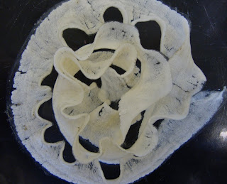
This is the brachiolaria larva of a starfish, Pisaster ochraceous,that I raised during the Embryology class. Larval anterior end is up, and you are looking at the ventral side. The brachiolaria is characterized by the presence of brachiolar arms and an adhesive disk, and the bottom image shows a close up of the pre-oral protuberance of the frontal lobe that bears these structures. The brachiolaria larva follows the bipinnaria larval stage in the forcipulates, a group of starfish.
The brachiolaria is the last larval stage in these asteroids. It is characterized by three brachiolar arms (the three stubby arm buds near the top of the larva in the first image and zoomed in on in the second image) which surround a central adhesive disk (the brown spot between the lower two brachiolar arms in both images). These appendages have sticky cells and are used to make contact with the substratum when the larva is competent to settle. Some other asteroids, that have lecithotrophic, or non-feeding development, skip the bipinnaria stage, and directly produce large yolky brachiolaria with three brachiolar arms and an adhesive disk also used for settlement and attachment.
Thursday, June 9, 2011
Wednesday, June 8, 2011
Spermatophore of Phoronopsis harmeri
The first image shows the spermatophore of a phoronid worm Phoronopsis harmeri. A spermatophore is a package that contains multiple sperm. Sperm is produced by males inside the body coelom, then released through the nephridiopores at the anterior end, and shaped into these packages by the special spermatophoral gland located inside the crown of tentacles (called the lophophore) which surrounds the mouth. The spermatophores of this species are round, about 300 micron in diameter, and equipped with a cork-screw shaped transparent “sail”. The sail is easily dislodged from the capsule.
The second picture shows two spermatozoa (or sperm). The sperm in this species is unusual in that it is V-shaped. The nucleus is in one arm of the “V”, and the flagellum forms the other arm, the acrosome is at the apex of the “V”. After the spermatophore is released it floats in the water and lands on the lophophore of a female. Once there, the spermatophore somehow makes its way into the tentacular coelom of the female, and eventually into the body coelom where internal fertilization occurs. Typically development is arrested until the female releases the fertilized eggs into the water. Occasionally development is initiated inside the female coelom, and one can find gastrulae or even more advanced developmental stages. See a post by Phillip Warner showing a 6-day old actinotroch larva of this species reared in the lab, and a post by Svetlana Maslakova showing an advanced actinotroch of this species collected from plankton.
Tuesday, June 7, 2011
Polar lobe in Nassarius fossatus
Nassarius fossatus is a marine snail. Snails, or gastropods, belong to the Spiralia - a large group of animals with spiral cleavage. Nassarius fossatus has unequal cleavage, which means that one of the cells at the two-cell and four-cell stage is larger than the others. The first four cells in a spiralian embryo are denoted as A, B, C and D. The D cell is the largest in unequal spiral cleavage. There are several mechanisms by which unequal cleavage can be accomplished. Nassarius does this via the so-called polar lobe, which is shown in these pictures. A polar lobe is an anucleated protuberance which forms at the vegetal pole during first, second, and sometimes subsequent cell divisions. It then fuses with one of the cells, making it larger than the others.
The top picture shows polar lobe formation during the first cell division. One can see two polar bodies. Polar bodies are the tiny sister cells of the oocyte which are produced during meiosis, contain discarded DNA and mark the animal pole of the embryo (up in the first three pictures). The opposite pole of the embryo is the vegetal pole. The two cells at the animal pole are the first two blastomeres. What looks like a third cell at the vegetal pole is the polar lobe, which at this stage is nearly completely cinched off from either blastomere. Subsequently the polar lobe fuses with one of the blastomeres (second picture from top), so that by the end of the first cell division one of the blastomeres (called CD) is noticeably larger than the AB cell (third picture from top). Polar lobe also forms at the second cell division (not shown). At the four-cell stage blastomere D is the largest, blastomere C is the second largest, while A and B cells are about the same size (bottom picture). The first three pictures are lateral views, while the bottom picture is a polar view. It is the first time I have heard of and observed unequal spiral cleavage, and I think it is remarkable. I also liked these eggs because the egg capsules they are laid in are very beautiful when viewed under the dissecting microscope (see picture by Janelle Urioste).
Gastropod Egg Masses
I was intrigued by the diversity of egg masses among marine gastropods (snails and slugs). In our Embryology class I observed three species with different habits of laying their eggs, namely, the snails Nucella emarginata, and Nassarius fossatus, and a nudibranch (or sea slug) called Diaulula sandiegensis. N. emarginata (also known as the “dog winkle”) and N. fossatus (also known as the “giant western dog whelk”) belong to the group of marine prosobranchs and produce egg capsules which are leathery or hard, and tend to be attached to various substrata in the environment. The top picture shows two egg capsules laid in our sea table by Nucella emarginata. These capsules are shaped like a wine goblet, and are opaque and hard, and each contains numerous eggs. The middle picture shows a single egg capsule of Nassarius. These are more or less transparent, so the eggs are easily visible through the wall of the capsule. When well fed, these scavenger snails will lay egg capsules attached to the walls of sea table, or the hose as close to the surface of the water as they can get. These egg capsules are beautifully sculptured. The bottom picture is of the egg mass laid in our sea table by the nudibranch Diaulula sandiegensis. Opisthobranchs, in general, lay their eggs in gelatinous ribbon-like masses. These masses are often loosely attached to the substratum, and tend to be only a few eggs thick.
Regeneration in bipinnaria II
In May I surgically bisected several bipinnaria larvae of the starfish Pisaster ochraceous. I made the cuts across the esophagus (anterior to the coeloms), separating the preoral lobe and the mouth from the rest of the larva (see pictures). I wanted to see if the fragments would indeed regenerate, as described in the literature.
The anterior and posterior fragments were cultured together and observed 13 days post-surgery. I observed two distinct morphotypes in the culturing vessel representing the anterior and posterior fragments. The first photo shows one of the posterior fragments 13 days after bisection. You can see that these fragments healed and regenerated the preoral lobe and the mouth. The fragments that I interpreted as being anterior, healed, but did not appear to regenerate 13 days after bisection (see bottom picture). This is surprising because bipinnaria larvae of the same species are apparently capable of regenerating their anterior ends under similar experimental conditions (Vickery et. al. 2002). I continued to monitor these fragments for several more weeks to see if the anterior ends would finally regenerate. As of six weeks post-surgery, I found two (out of original 15) anterior fragments that did not appear to regenerate.
Vickery, M. S., Vickery, M. C. L., McClintock, J. B. 2002. Morphogenesis and Organogenesis in the Regenerating Planktotrophic Larvae of Asteroids and Echinoids. Biol. Bull. 203: 121–133
Monday, June 6, 2011
Pseudocyphonautes of Flustrellidra
These pictures are of the bryozoan Flustrellidra coniculata and its pseudocyphonautes larva. The pseudocyphonautes resembles the planktotrophic cyphonautes larva of some bryozoans in that it has a bivalve shell. But it is lecithotrophic (non-feeding) and is brooded. The top picture shows the bean-shaped pseudocyphonautes larva using a dark-field technique. One can distinguish the cilia of the corona ciliata along one edge of the larva. One can also see a ridge on the opposite side which corresponds to the shell margin. The clear outer coating is the shell that protects the larva’s interior structures.
The middle picture shows a small section of the colony (with zooid lophophores retracted). The three white oval shapes out of focus are the larvae which I have dissected. Just beneath the surface of the colony one can see a few yellowish-white masses. These are the ovicells, specialized zooids for brooding eggs and larvae. I extracted the pseudocyphonautes larvae by puncturing the ovicells with a pair of fine forceps and squeezing gently to push them through the hole.
The bottom picture shows a complete Flustrellidra colony, with my palm for scale. This is the largest upright bryozoan colony I have ever seen. It does not look like a bryozoan at all. In the field I would have easily mistook this animal for some sort of alga or a sponge!
Thursday, June 2, 2011
Brooded ophiuroid juveniles
Amphopholis squamata is a common small intertidal ophiuroid (brittle star) in the Pacific NorthWest. Unlike many larger species of ophiuroids which produce large numbers of eggs, free-spawn them and have pelagic development, this small species produces few eggs at a time and broods its young in specialized pouches, called the genital bursa, located in the central disc. The juveniles may be removed from the brooding adult by opening the genital bursa from the oral side. The juveniles dissected from a single adult are often at different developmental stages and of different size. They may range from fertilized egg to a juvenile ready for self-sufficiency. The different stages may be found within a single brooding pouch. These are dark-field photomicrographs of two juveniles dissected from a single adult taken at the same magnification. The top picture is of a small juvenile that was still connected to the brood pouch of the adult brittle star, so that it may receive necessary nutrients for development. Because the embryos receive nutrition from the mother, they grow inside the brood pouches. The bottom picture shows a larger juvenile which has developed arms.
Raising larval cultures
In this Embryology class we raise embryos and larvae to gain a better understanding of how organisms develop. It is not hard to raise larvae, but it must be done with care. All of the species we work with are local marine invertebrates, collected either by us during the class field trips, or by our instructor ahead of time. The first step is to procure gametes, and the technique varies by species. Once we have fertilized eggs, we culture them in filtered sea water in clear glass custard bowls that hold 100-200 ml. The bowls are set in a sea table with flowing sea water, so the cultures are kept at ambient sea temperature. We label cultures using clothespins and colored tape (top picture).
Every other day the water must be exchanged using reverse filtration. A small plastic beaker with a mesh bottom (mesh size of 50-100 microns) is set in the culture bowl, and water is removed with a turkey baster, while the embryos or larvae remain in the bowl as you can see one of the students do in the middle picture. Bacteria and detritus go through the mesh together with the water. You must choose an appropriate mesh size so that the larvae are not going through it and make sure you leave a small amount of water on the bottom with larvae, so they are not crushed.
The next step is to transfer your larvae into a clean bowl and fill with filtered seawater. Some of our cultures need food to grow and develop. We have been feeding them a mixture of two kinds of unicellular algae, Rhodomonas lens (looks red) and Dunaliella tercioleta (looks green), which our teaching assistant grows in the lab in 0.5 liter glass flasks shown on the bottom picture.