Tuesday, March 26, 2013

Development of a hoplonemertean


On February 4, 2013, I did a plankton tow off a dock in Charleston, OR and found a few embryos like the one pictured here. The embryo was about 180 microns across, and was surrounded by an inner chorion and an outer chorion about 360 microns in diameter. I kept the embryos, and after two days discovered that they turned into planuliform larvae, hatched from their chorions and were swimming around in the dish.

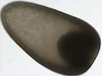
The planuliform larva is uniformly ciliated and has a prominent bundle of cilia called the apical tuft at the anterior end (12 o’clock on the second picture). Such larvae are found in nemerteans (phylum Nemertea, a.k.a. ribbon worms) from the orders Hoplonemertea and Palaeonemertea. 

After a few more days these larvae developed features that allowed me to identify them as belonging to the order Hoplonemertea (e.g. several pairs of subepidermal eyes visible on the third picture). This picture shows an 11-day old individual. At this point they mostly crawled on the bottom of the dish, and had developed many of the adult structures, such as the brain and proboscis, so they can be considered juveniles rather than larvae. Hoplonemertean metamorphosis (the transition from planktonic larva to benthic juvenile) is inconspicuous. The transition from swimming to crawling is accompanied by changes in the epidermis. Apparently many hoplonemerteans replace the larval epidermis composed of large, ciliated, cleavage-arrested cells with intercalating smaller cells of the definitive epidermis (Maslakova and von Döhren, 2009).

To witness this epidermal transition, I fixed some of my specimens in paraformaldehyde (with a touch of gluteraldehyde) and stained them with fluorescent phalloidin to visualize the outlines of the epidermal cells using a confocal microscope.

The first confocal image shows a 2-day old hoplonemertean planuliform larva – the same age as the live larva pictured above. You will notice that large cells dominate the epidermis, but small cells are visible in between. These small cells are the intercalating cells of the juvenile epidermis. The bottom image is a 4-day old larva, and you can see that the large cells of the larval epidermis are farther apart from each other. The small cells of the juvenile epidermis occupy more space in between. Eventually, cells of the larval epidermis will be either resorbed or sloughed off, leaving the cells of the definitive epidermis to cover the entire surface. 

Maslakova SA, von Döhren J. (2009) Larval development with transitory epidermis in Paranemertes peregrina and other hoplonemerteans. Biol Bull 216: 273-292

Observations of a magelonid nectochaete larva


In a plankton tow taken in Charleston, OR on February 20, 2013, I found several polychaete nectochaete larvae of Magelona sp., which I was able to identify by their long anterior tentacles. The tentacles apparently develop as extensions of the prototroch (larval ciliary band) and are suspected to be used in locomotion and feeding  (Wilson 1982). I noticed the tentacles were relaxed and extended when the larva was hanging suspended in the water column, but during rapid sinusoidal movements of the body, the larva coiled up its tentacles and kept them close to the body.

This picture is a close up of one of the tentacles. You will notice that they are covered by papillae. The papillae are present along the entire length of the tentacle but only on the dorsal side. Pairs of papillae appear to be at right angles to each other as they each emerge from the surface of the tentacle. It is suspected that the papillae may serve a sensory function by detecting vibrations in the surrounding water (Jones 1968).


This is a close up of the larval mouth (ventral view) - note its distinct triangular shape. Two eyespots are also noticeable near the anterior end of the larva. Toward the posterior (down), bundles of bristles emerge from paired chaetal sacs on each segment of the body.

Wilson DP. 1982. The larval development of three species of Magelona (Polychaeta) from localities near Plymouth. J Mar Biol Ass UK 62: 385-401

Jones ML. 1968. On the morphology, feeding, and behavior of Magelona sp. Biol Bull 134(2): 272-297.


Feeding mechanism of the actinotroch larva


This picture shows an unidentified actinotroch larva of a phoronid worm that I caught in a plankton tow taken off a dock in Charleston, OR on February 20, 2013. The photo is a lateral view, and the anterior end of the larva is up. Actinotroch larvae have an anterior hood that covers the mouth (a lobe-like structure seen at upper left) as well as a crown of tentacles located posterior to the mouth. You can see that the tentacles are longer ventrally and shorter dorsally. This is because tentacle pairs are added progressively with new tentacles forming mid-dorsally, so the mid-ventral tentacles are the oldest. The number of tentacles varies between species. This larva had ~ 20 tentacles. Tentacles are involved in feeding. A posterior ciliated band, called the telotroch (at about 6 o-clock), helps in locomotion.


The second picture is a close up of the tentacles showing the cilia. The tentacles are covered by motile cilia, which create the water flow, but also bear a row of non-motile latero-frontal cilia. These stiff latero-frontal cilia apparently serve as a mechanical sieve, detaining food particles as they flow past the larva (Riisgård, 2002). Once caught on the upstream side of the tentacle, a food particle can be transported up to the mouth by the beating frontal cilia (Riisgård 2002), a tentacle flick (Strathmann and Bone 1997), or rapid lifting of the preoral hood which creates negative pressure (Strathmann and Bone 1997).

I gave my actinotroch some unicellular algae Rhodomonas (from a lab culture), which is a good food for many ciliated marine invertebrate larvae. This picture was taken a day after food was added, and it appears that the larvae fed on Rhodomonas, judging from the accumulation of the reddish pigment (the color of Rhodomonas) in the stomach.

Riisgård HU. 2002. Methods of ciliary filter feeding in adult Phoronis muelleri (phylum Phoronida) and its actinotroch larva. Mar Biol 141: 75-87.


Strathmann RR and Bone Q. 1997. Ciliary feeding assisted by suction from the muscular oral hood of phoronid larvae. Biol Bull 193: 153-162.

Wednesday, February 27, 2013

Secrets of a mollusk egg


I found a few of these interesting embryos in a plankton tow taken off a dock in Charleston, OR in January 2013. Under a dissecting microscope, I could see the embryo itself was greenish and about 250 microns across, but it seemed to be surrounded by a halo several hundred microns in diameter. When I sucked the embryo into my pipet, I could see the “halo” contacting the inside walls of the pipet. As it turns out, the “halo” is a jelly layer, a characteristic of some embryos that, among other things, helps protect it from microbes (Hellberg et al. 2012). Oregon Institute of Marine Biology research scientist George von Dassow identified the specimen as a gastropod mollusk (snail) embryo.

A closer look under a compound microscope revealed many fascinating things about this embryo. In the first photo, you’ll notice two small, clear cells at about 4 o'clock. These cells, called polar bodies because they mark the animal pole, are essentially the waste products of meiosis. In many invertebrate phyla, meiosis II is not completed until after fertilization, so the resulting polar bodies remain inside the chorion (the membrane surrounding the embryo inside the jelly).

In the next photo, you should notice two things: first, there seems to be a hole in the chorion at about 10 o'clock (a penetration point of sperm), and second, a sperm is hanging out just inside the chorion at about 7 o'clock. Other similar penetration points were visible in other parts of the chorion, indicating that multiple sperm were able to enter the chorion but did not fertilize the egg (because the embryo looks healthy). Polyspermy is actually a big problem for invertebrates, especially those that are free-spawning and have external fertilization (Franke et al. 2002). Polyspermy causes morphological abnormalities in development, so complex physiological mechanisms exist to prevent it. A close coupling exists between the species-specific surface proteins on sperm (e.g. lysin) and egg (vitellin envelope receptor for lysin), so interspecific hybridization is also prevented (Hellberg et al. 2012).

After a few days, the embryo had developed into a gastropod veliger larva about 400 microns long. The reticulated pattern on its shell suggests this veliger belongs to the turban snail genus Calliostoma, of which there are 5 species in Oregon (Goddard 2001). The larva uses the compound cilia of its velum (ciliated structure at 4 o'clock) to feed and move throught the water column.


Franke ES, Babcock RC, Styan CA. (2002) Sexual conflict and polyspermy under sperm-limited conditions: In situ evidence from field simulations with the free-spawning marine echinoid Evechinus chloroticus. American Naturalist 160(4): 485-496

Goddard JHR. 2001. Mollusca: Gastropoda. In: An Identification Guide to Marine Larval Invertebrates of the Pacific Northwest. Edited by Alan Shanks. OSU Press, Corvallis.

Hellberg ME, Dennis AB, Arbour-Reily A, Aagaard JE, Swanson WJ. (2012) The Tegula tango: A coevolutionary dance of interacting, positively selected sperm and egg proteins. Evolution 66(6): 1681-1694

Thursday, December 13, 2012

Confirmed identity of wild-caught planktonic larvae

We recently confirmed the identity of two planktonic larvae that were each the subject of blog posts last spring.  Both larvae were cryopreserved after photographs were acquired and their DNA was extracted at the Oregon Institute of Marine Biology by a student in Svetlana Maslakova’s Marine Molecular Biology class this fall.

Nephtys sp. metatrochophore 

Metatrochophore larvae like this were found in April and May and we speculated that they were larvae of the polychaete genus Nephtys due to the red pigment bands along the prototroch and pygidium as well as the bright blue pigment lining the stomach which you can recall from this photograph (for full blog post see Nephtys sp. metatrochophore by Terra Hiebert).  

Jay Bowles, a student in the Marine Molecular Biology class, successfully amplified the 16S rDNA barcoding gene region for this larva with universal primers (Palumbi et al. 1991).  Using NCBI BLAST, we compared this sequence to those in GenBank and found that this larva is, in fact, the larva of Nepthys as its sequence matched other species within this genus with 97% sequence similarity.  The species level identify of this larva remains to be determined, as sequences did not match exactly, but we’ve confirmed our previous identification based on larval morphology.  

Holothuroid doliolaria 

I found this larva in a plankton sample taken from the Charleston marina docks on May 17th.  We speculated that it may be the larva of a sea cucumber (class Holothuroidea) because of its unique barrel-like shape and transverse ciliated bands (for full blog post see Holothuroid doliolaria by Terra Hiebert).  

Jay successfully amplified the Cytochrome Oxidase Subunit I barcoding gene region from this larva, also using universal primers (Folmer et al. 1994).  Unlike the Nephtys sequence mentioned above, the sequence we acquired from this larva did not have a close match to any sequences in GenBank.  The closest match (87% similarity) was to the sea cucumber Acaudina molpadioides. Richard Emlet (pers. comm.) suspected that our larva may belong to the local holothuroid species, Paracaudina chilensis (rat-tail sea cucumber), based on its morphology and development, so we compared the larval COI sequence to that obtained from P. chilensis by Gustav Paulay (University of Florida, Florida Museum of Natural History).  The two sequences had only 4 mismatches over the 490 base pair region of overlap (0.8% divergence).  This level of divergence is low enough that we can be confident that our doliolaria larva belongs to P. chilensis.  

While pursuing the identification of this larva with DNA sequence data, we realized that we misinterpreted the madreporite as ossicles in the polarized light image you can see here.  Calcareous ossicles are found in sea cucumbers and are used for structure and support.  However,  as we now understand, they usually do not develop until much later stages (Richard Emlet, pers. comm.).  The madreporite, on the other hand, is a small calcareous disc that connects the water-vascular system to the outside, and is used to regulate internal water content in all echinoderms, including sea cucumbers. Madreporites have been documented in young Paracaudina sp. larvae (Richard Emlet, unpublished) and resemble the brighter spicule on the right in the photograph shown here.  In addition to the madreporite, the water vascular system of echinoderms includes another calcareous structure called the ring canal that helps to move water throughout the body.  We speculate that the ring canal can also be seen in this image and is the larger structure to the left of the madreporite. 

Folmer O, Black M, Hoeh W, Lutz R, Vrijenhoek R. (1994) DNA primers for amplification of mitochondrial cytochrome c oxidase subunit I from diverse metazoan invertebrates. Mol Mar Biol Biotech 3:294-299 

Palumbi S, Martin A, Romano S, McMillan WO, Stice L, Grabowski G. (1991) The simple fools guide to PCR Version 2.0.  Honolulu, HI: Department of Zoology Kewalo Marine Laboratory, University of Hawaii

Thursday, August 23, 2012

MOCNESS


Last month I participated in an oceanographic research cruise near the Barbados Accretionary Prism. My job was to help with the MOCNESS (Multiple Opening/Closing Net and Environmental Sensing System), which is a net system used to sample plankton. The MOCNESS can collect independent plankton samples at specific depth intervals during a single deployment, this is accomplished by independently opening and closing the nets.

The MOCNESS has a rectangular frame that houses the environmental sensing system and controls the nine nets (MOCNESS can have between 6 and 20 nets). The cable connects the ship and MOCNESS and gives scientists real-time data so instrument adjustments can be made during sampling. The sensors were used to collect salinity, temperature, depth, and water flow measurements. We used 153 µm mesh nets and the speed of the MOCNESS through the water averaged 10m/min so that the small ciliated larvae would not get damaged. 

The pictures are of the MOCNESS after it had been recovered. The first image (taken by Svetlana Maslakova) is the science team transferring the samples from the cod ends to containers for sorting. The second image (taken by Svetlana Maslakova) is of myself and another graduate student setting up the nets in preparation for the next tow. The third image is a sample that was collected in one of the cod ends. We conducted nine MOCNESS tows, depths of the samples ranged from the surface to 4500 meters. These tows will allow us to look at the vertical distribution of deep sea larvae. 

Wiebe, P. H., K. H. Burt, S. H. Boyd and A. W. Morton. 1976. A multiple opening/closing net and environmental sensing system for sampling zooplankton. Journal of Marine Research 34:313-326

Wiebe, P. H., A. W. Morton, A. M. Bradley, R. H. Backus, J. E. Craddock, V. Barber, T. J. Cowles and G. R. Flierl. 1985. New developments in the MOCNESS, an apparatus for sampling zooplankton and micronekton. Marine Biology 87:313-323

Thursday, June 28, 2012

Inarticulate Brachiopod Larvae

This June I participated in an oceanographic research cruise to Barbados. One of the goals of this expedition was to collect planktonic larvae of benthic marine invertebrates using a MOCNESS device. Among other things, we found larvae of inarticulate brachiopods (phylum Brachiopoda; class Inarticulata) pictured here. Brachiopods resemble bivalve molluscs (e.g. clams and mussels), but have dorsal and ventral valves rather than left and right. The valves are held together with muscles in inarticulate brachiopods, while those of articulate brachiopods are hinged. Brachiopods, as a phylum, are also characterized by the lophophore (a crown of tentacles surrounding the mouth). We collected two different kinds of inarticulate brachiopod larvae.

Larvae of the first kind were collected from relatively shallow depths (900-0 m). In this image, one can see the large circular larval shell valves, as well as the embryonic shell (the half circle at the posterior portion of the larval shell, tinged blue). The lophophoral tentacles are retracted in the first image, but extended in the second.

These tentacles form progressively during the larval life from short tentacle buds on either side of a longer unpaired anterior median tentacle (at about 12 o’clock on the second image). Consequently, the number of tentacles reflects larval age. This larva has 10 pairs of tentacles. In some species, a lophophore with this many tentacles indicates larval competence for settlement (Pennington and Stricker, 2002). One can also see that the tentacles of the lophophore are ciliated. Cilia are used for larval feeding and swimming (Rudwick 1970). This particular type of larva is produced by members of the superfamily Lingulacea which contains only two extant genera: Lingula and Glottidia (Pennington and Stricker 2002).

Larvae of another kind were collected from deeper waters (1600-900 m). Characteristically, they lack an embryonic shell, and have a pair of larval chaetae. Presence of chaetae in this larva suggests that it belongs to the inarticulate superfamily Discinacea (Pennington and Stricker 2002). Chaetae are usually thought of as a feature unique to annelid worms, but some brachiopod larvae and adults also have chaetae, which are similar structurally and developmentally to those of annelids. 

Pennington, J T and S A Stricker. 2002. Phylum Brachiopoda. In: Atlas of Marine Invertebrate Larvae. Edited by Craig M Young. Academic Press. 

Rudwick, M J S. 1970. Living and Fossil Brachiopods. Hutchinson & Co, London.

Monday, June 4, 2012

Fan worm larva

On May 10th, 2012 I waded through the calm waters of Middle Cove at Cape Arago State Park, trailing a 153μm plankton net behind me.  While sorting through the plankton I found a polychaete larva from the family Serpulidae, subfamily Spirorbinae.  Serpulids belong to an order of annelids, Sabellida, commonly known as fan worms.  Adult Spirorbins live in small calcareous tubes approximately 2-5 mm in diameter typically attached to undersides of intertidal rocks. Adults are hermaphroditic and brood embryos inside their tube, then release lecithotrophic larvae that are only briefly planktonic (Strathmann 1987).

This metatrochophore larva has a prominent ciliated band anterior to the mouth, called the prototroch, which is used for swimming. At the anterior (up) is the apical tuft, and at the posterior end there is another cilary band, called telotroch. What helped us to identify this larva is the prominent collar located posterior to the prototroch (Kupriyanova et al. 2001). The collar is the widest portion of the larval body. This larva had  5-6 segments posterior to the collar, and three pairs of ocelli. 

To my surprise, when I went to photograph the larva a few days later, I could not find it at first.  No longer content with the planktonic life, it had settled on the bottom of the culture dish and built a calcareous tube (left). The tube is smooth, non transparent, and coils to the right (dextral). The characteristics of the tube helped us to further classify this specimen as likely belonging to the genus Circeis (Blake and Ruff 2007). 

You can see on the subsequent photograph that it got quite a bit longer in 4 days. The juvenile worm is caught peaking out of it’s tube in the first picture. One can discern a radiolar crown of tentacles used for feeding and respiration (Blake and Ruff 2007), red eyespots, and an operculum.  When disturbed the animal quickly retreats back into its tube, and shuts the operculum (below).

Blake JA, and Ruff RE.  2007. Polychaeta.  In: The Light and Smith Manual:  Intertidal Invertebrates from Central California to Oregon 4th Edition.  Edited by James Carlton. University of California Press, Berkeley.  

Kupriyanova, E.K., E. Nishi, H.A. ten Hove, & A.V. Rzhavsky 2001. A review of life history in serpulimorph polychaetes: ecological and evolutionary perspectives. Oceanography and Marine Biology: an Annual Review 39:1-101.

Strathmann, Megumi F. 1987.  Phylum Annelida, class Polychaeta. In: Reproduction and Development of Marine Invertebrates of the Northern Pacific Coast.  United States: University of Washington Press. 

Sunday, June 3, 2012

Development of the sea cucumber Cucumaria miniata

This series of images illustrates the development of a sea cucumber Cucumaria miniata. I collected adults of C. miniata from Lighthouse beach in Charleston, Oregon on April 24 2012 hoping to obtain gametes from natural spawning because this species is known to reproduce from mid-March to late April. I kept the adults in a sea table with flowing sea water at ambient temperature and crossed my fingers. On April 28 2012, two males and three females spawned. The adults were separated before spawning, so I collected the gametes and fertilized the eggs. Cucumaria miniata is considered a direct-developer. Even though it has a swimming larva, called doliolaria, this larva does not feed in the plankton. In contrast, some other sea cucumbers have a feeding auricularia larva, which eventually metamorphoses into a doliolaria stage. As is typical for direct developing sea cucumbers, C. miniata has large yolky eggs (500 µm in diameter and bright green in this species). A few hours after fertilization I observed early cleavage – a four cell stage is pictured here.

The second picture shows a 60-hour old early doliolaria larva of C. miniata – the preoral lobe (right) is more opaque compared to the posterior end of the larva (left). Although these do not feed they do have a vestige of a gut. Early on the larva is uniformly ciliated, but the advanced doliolaria larva of this species swims using three transvers ciliary bands (not shown).

By day seven of development one can see there are five-primary tentacles protruding ventrally toward the anterior end of the larva (about six o'clock on the image on the left). The five primary tentacles surround the mouth. At this stage the individual is referred to as a pentactula. The pentactula of this species also has two primary podia (one clearly visible on the image to the left) emerging from little pores (called podial pits) at the posterior end.  

The bottom image shows several of the many calcareous spicules in the pentactula. After two weeks of planktonic life the larvae (as pentactulae) settle on the undersides of rocks near conspecific adults.

Sewell, M. and McEuen, F. Phylum Echinodermata: Holothuroidea. In: Atlas of Marine Invertebrate Larvae. Edited by Craig M Young. Academic Press.

McEuen, F. Phylum Echinodermata, Class Holothuroidea. In: Reproduction and Development of Marine Invertebrates of the Northern Pacific Coast. Edited by Megumi F Strathmann. University of Washington Press.

Genital plate formation in echinopluteus larva

Indirect development often involves a major change in body plan. The larva of the purple sea urchin, Strongylocentrotus purpuratus, has bilateral symmetry, and the adult has 5-fold radial symmetry. As the echinopluteus larva approaches metamorphosis clues to the body-axis shift start to appear. The first image shows a lateral view of the larva. The two vertical white lines are calcareous rods that support the arms of the larva.  They appear to glow because I used polarized light microscopy. Forming around each rod is a honeycomb-like structure which will become one of the genital plates in the adult. After metamorphosis, these plates are located on the aboral side of the adult (the side opposite the mouth) and surround the mouth. They are called genital plates because the gonads (testes or ovaries) open to the outside via little holes (called gonopores) in these plates. The endoskeleton of adult urchin, called the test, is made up of many such closely fitted calcareous plates.

The second picture is a close-up aboral view of the test of an adult urchin. Surrounding the large hole in the center (the anus) are the apical plates. You can also see five smaller openings, the gonopores, in the genital plates. Starting at 12 o’clock is the genital plate 2 or G2. This much larger plate perforated by a large number of small pores is called the madreporite; it connects the water-vascular system of the urchin to the outside. Clockwise from G2 are the other four genital plates in the following order: G3, G4, G5, and finally G1. I think the left and right genital plates on the larval image above correspond to G5 and G3 respectively (Emlet, 1985).

Emlet, R. B. 1985. Crystal Axes in Recent and Fossil Adult Echinoids Indicate Trophic Mode in Larval Development. Science 230: 937-940.

Saturday, June 2, 2012

Müller’s larva

On May 3rd and 4th 2012, I performed plankton tows at the South Cove of Cape Arago, near Charleston, OR as the tide was coming in.  I placed a 153μm plankton net into a channel of water flowing between smooth rocks. In this manner I was able to obtain interesting samples, which included, among other things, many specimens of Müller’s larva pictured here. 

Johannes Müller (1801-1858), a German physiologist and the inventor of the plankton net, first described larval forms of many phyla including Platyhelminthes, or flatworms, whose planktonic larvae retain his name (Smith et al. 2002). The term “Müller’s larva” is used to describe planktonic larvae in the Polycladida order of the class Turbellaria. Müller’s larva is ciliated and characterized by several paired and unpaired lobes (left). The lobes bear cilia that are longer than cilia on the rest of the body. At the anterior (up) and posterior (down) end of the larva there are tufts of longer cilia (apical and caudal, respectively). The apical tuft originates from the apical organ, a sensory structure that is associated with the central nervous system (Rawlinson 2010).

When I collected these larvae they were fairly uniform in size approximately 180 μm long by 80 μm wide. Their shape was simliar, with lobes present in all of the specimens.  When I observed them two weeks later some had lost their characteristic lobes. The second photo shows a larva where the lobes are present, but do not protrude much.  Müller’s larvae may also have several pairs of eyes.  The two areas of black pigmentation at the anterior portion are a cerebral eye and an epidermal eye, which is closer to the apical tuft. 

The last photo (left) shows a, presumably, more advanced larva that has completely resorbed its lobes and has begun to take the characteristic shape of an adult flatworm. In this larva you can clearly see the two cerebral eyes, with one of the epidermal eyes lower and to the right. When observed two weeks later this larva had not noticeably changed in shape or form. Little is known about the development of Müller’s larva, and it would be interesting to investigate the mechanisms driving these morphological changes.

Smith NF, Johnson KB and Young CY. 2002. Platyhelminthes. In: Atlas of Marine Invertebrate Larvae. Edited by C. M. Young. Academic Press. New York.

Rawlinson KA. 2010. Embryonic and post-embryonic development of the polyclad flatworm Maritigrella crozieri; implications for the evolution of spiralian history traits. Frontiers in Zoology. 7:12

Friday, June 1, 2012

Chiton development


This picture shows a chiton egg which I collected from the plankton on February 8th, 2012, off of a dock in Charleston, OR. Chitons are marine mollusks characterized by a shell made up of eight separate plates. Chiton shell plates washed up on the beach are often referred to as “butterfly shells.” Chiton eggs are very distinctive because they are surrounded by a thick and often ornate hull (as seen in this picture). The hull has been found to reduce the sinking rate of the egg as well as to focus sperm to specific regions on the egg surface (Buckland-Nicks, 1993) . 

This is a trochophore larva that hatched out of a chiton egg. Chiton trochophores have a long apical tuft and an equatorial ciliary band called a prototroch, which you can see on this picture. Chiton trochophores are different from any other kind of trochophore because their two eyes are located posterior to the prototroch (instead of anterior). After a few days of development these larvae begin to form shell plates. 

This picture shows seven transverse bands on the dorsal side of the larva posterior to the prototroch. These bands delineate the boundaries of the future shell plates; the eighth plate appears later in development (anterior to the prototroch). The dark band is part of the mineral skirt, which can be seen more clearly in the photo below. The foot and mouth of the chiton is developing on the ventral surface. 

I took this picture using polarized light to show the mineral spicules in the epidermis surrounding the shell (this area is called the girdle) and the seven initial shell plates which are beginning to form.   

Buckland-Nicks, J. 1993. Hull capsules of chiton eggs: parachute structures and sperm-focusing devices? Biological Bulletin. 184: 269-276. 

Thursday, May 31, 2012

Larva of Polygordius sp.

Although this might look like a picture of a flying saucer it is actually a lateral view of a trochophore larva of a polychaete from the genus Polygordius. This larva (and many others like it) were collected from the plankton in Charleston, Oregon on May 10th. The Polygordius trochophore was the first trochophore larva described in the literature (Hatschek 1878), even though it is quite unusual, as far as trochophore larvae go (Rouse 1999). Trochophore larvae, in general, are characterized by having an equatorial ciliary band, called the prototroch. You can clearly see the prototroch in the first picture as a yellow band that divides the larva into two regions - the upper episphere, and the lower hyposphere. The unusual thing about this trochophore, also known as “endolarva” (Woltereck 1904), is that the segmented juvenile body develops tucked inside the spherical larval body. The larva pictured here has a well developed juvenile body inside the transparent hyposphere. The two red spots at the apex of the episphere  are the eyes.  The greenish/yellow ballon inside the larval sphere is the stomach. Polygordius larvae have a through gut and feed in the plankton. 

This picture offers a view of the larval apical organ located at the apex of the episphere. The two eyes which are part of it appear black in transmitted light. The apical organ also contains two clear vesicles, which look like statocysts. Statocysts are balance organs found in some larval and adult marine invertebrates. 

Polygordius trochophore is one of the few polychaete larvae that have a catastrophic metamorphosis. I was fortunate enough to witness this process in the lab, and took a picture, as the larva was undergoing metamorphosis (left).  The orange-yellow band at the anterior end (up) is the prototroch. The process of metamorphosis begins with the juvenile suddenly extending out of the hyposphere. The larval body (including the prototroch) is then resorbed.  In this larva the process was more gradual than in some others of this genus - the episphere was resorbed gradually over the course of a few days, and even after a week one could still detect the remnants of the prototroch, even as the now juvenile worm crawled around the bottom of the culture bowl. 

Hatschek B. 1878. Studien über Entwicklungsgeschichte der Anneliden. Ein Beitrag zur Morphologie der Bilaterien. Arbeiten aus dem Zoologischen Institute der Universität Wien und der Zoologischen Station in Triest. 1: 277–404. 

Rouse G. 1999. Trochophore concepts: ciliary bands and the evolution of larvae in spiralian Metazoa. Biological Journal of the Linnean Society. 66: 411–464. 

Woltereck R. 1904. Beiträge zur praktischen Analyse der Polygordius-Entwicklung nach dem “Nordsee”-und dem “Mittelmeertypus”-Typus. I. Die für beide Typen gleichverlaufende Entiwicklungsabschnitt: Vorm E ibis zum jungsten Trochophore-Stadium. Arch. Entw.Mech. Org. 18: 377-403.

Monday, May 28, 2012

Holothuroid doliolaria

I found this gastrula in a plankton sample taken from the Charleston marina docks on May 17th.  We speculated that it may be a sea cucumber (class Holothuroidea; phylum Echinodermata) because of its unusual shape.  One can see in this picture that the gastrula is longer than wide and tapers toward the animal pole (upper left).  In holothuroids, gastrulation begins by invagination whereby cells at the vegetal pole (bottom right) fold into the blastocoel (the cavity within the embryo) as a layer, rather than ingress individually (McEuen 1987).  One can see these invaginating cells in this gastrula as an opaque area.  This embryo later confirmed our suspicions and developed into a lecithotrophic (non-feeding) doliolaria (planktonic larva of some holothuroids). 

Doliolariae are shaped like a barrel (hence the name) and are ciliated. Cilia may cover the entire surface (uniform ciliation) or form 2-5 discrete transverse bands.   Ciliated bands often develop from an initially uniformly ciliated area of epidermis (Miller 2001).  On May  23rd (left) two ciliary bands were present in this larva in addition to a uniformly ciliated anterior area (left on this image). 

By May 25th, the uniformly ciliated region had developed into yet another discrete band of cilia (three in total), seen in the third photograph.The duration of the doliolaria larval stage is species specific, ranging from six to thirteen days in the local species with described development (Miller 2001). The end of planktonic period is marked by the protrusion of five primary tentacles from the larval vestibule.  One can see several curved projections in the larval epidermis (upper left in the third image) through which these tentacles will eventually emerge. At that point the larva will be called a pentactula.

Although sea cucumbers are relatively soft bodied, internal calcareous ossicles are used for structure and support.  In this polarized light image, one can see a small ossicle as well as a larger ring of ossicles that will surround the pharynx in the adult sea cucumber (Ruppert et al. 2004). 

Miller, B. 2001. Echinodermata. In: An Identification Guide to the Larval Marine Invertebrates of the Pacific Northwest. Edited by Alan Shanks. OSU Press, Corvallis.

McEuen, SF.  1987.  Echinodermata: Holothuroidea.  In: Reproductive and Development of Marine Invertebrates of the Northern Pacific Coast by Megumi Strathmann.  University of Washington Press, Seattle. 

Ruppert, E. E., R. S. Fox and R. D. Barnes.  2004.  Invertebrate Zoology: A Functional Evolutionary Approach.  Brooks Cole, Belmont, CA.

Identifying crustacean nauplii

The first picture is that of a copepod nauplius from a plankton tow collected at the mouth of Coos Bay, OR on Saturday, May 5th. The nauplius is the first larval stage of many crustaceans (although many species pass through this stage while still enclosed in the egg capsule). Nauplii are characterized by having three pairs of appendages: a pair of uniramous (not forked) antennules, and a pair of antennae and manidibles, both of which are biramous (i.e. forked). Nauplii are some of the most common organisms one encounters while sorting plankton samples. In Coos Bay we often encounter the nauplii of both barnacles and copepods.

In the second picture we see the nauplius of a barnacle.  Typically barnacles go through four to six naupliar stages depending on species. The distinguishing characteristic of a barnacle nauplius is the pair of fronto-lateral horns at the anterior end. Copepod nauplii do not have any such horns. Being able to distinguish between the two kinds of nauplii is important because both have a biphasic life cycle, but copepods are holopelagic (meaning they spend the entire life cycle in the water column) whereas barnacles are benthopelagic (adults are benthic, while larvae are planktonic) so different ecological questions can be addressed by studying these two kinds of organisms.

After the nauplius barnacles go through an additional larval stage called the cyprid (left). The final nauplius stage molts into the cyrpid stage, which is non-feeding. Cyprid larva has a pair of large appendages at the anterior end (left side of this photo) called antennae  - one can see these sticking out from the carapace. The cyprid will use these to “walk” around on the substrate while  looking for an ideal spot to settle. Once it finds a spot it will cement itself to the substrate and undergo metamorphosis into an adult (permanently attached) stage.